
Rhinovirus, member of the genus Enterovirus family Picornaviridae, is a positive-sense, single-stranded-RNA (ssRNA) virus of approximately 7.2 kb. Currently, more than 160 sero-/genotypes have been described and classified into three species, RV-A, RV-B and RV-C. Sequencing and serologic methods have defined approximately 83 HRV-A types, 32 HRV-B types, and 55 HRV-C types with potentially as many as 150 to 170 serological distinct HRV types in circulation. Human rhinoviruses (HRVs) were first discovered in the 1950s in an effort to identify the etiology of the common cold. While once thought to cause relatively benign upper respiratory tract illness, HRVs also are important cause of otitis media and sinusitis and can precipitate exacerbations of asthma in children. Rhinovirus infection may play role in other illnesses that result in hospitalization of young children. Transmission of HRVs occurs primarily between individuals by either direct contact, contact with fomites, or aerosols.
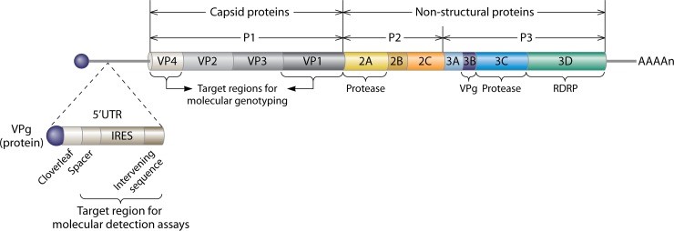
HRV is a non-enveloped virus with a positive-sense single-stranded RNA that encodes 11 proteins. The viral polyprotein is divided into a P1 region, which encodes the capsid proteins VP1, VP2, VP3, and VP4, and the P2 and P3 regions, which include proteins 2APro, 2B, 2C, 3A, 3B (VPg), 3CPro, and 3DPol. Four proteins, VP1, VP2, VP3, and VP4, make up the viral capsid that encases the RNA genome, while the remaining nonstructural proteins are involved in viral replication and subsequent assembly. Antigenic variation among HRV types is derived from variations in the exposed surface of VP1, VP2, and VP3, while embedded VP4 is responsible for RNA packaging during assembly. Compared to the rest of the HRV genome, the capsid proteins exhibit a high degree of heterogeneity resulting in a wide range of antigenic diversity. There are 60 copies each of the four capsid proteins, giving the virion an icosahedral structure, with a canyon in VP1 that serves as the site of attachment to cell surface receptors.
- Capsid protein VP1 forms an icosahedral capsid of pseudo T=3 symmetry with capsid proteins VP2 and VP3. VP1 interacts with host cell receptor to provide virion attachment to target host cells. VP1 N-terminus (that contains an amphipathic alpha-helix) and VP4 are externalized, they shape a pore in the host membrane through which viral genome is translocated to host cell cytoplasm.
- Capsid protein VP2 forms an icosahedral capsid of pseudo T=3 symmetry with capsid proteins VP1 and VP3.
- Capsid protein VP3 forms an icosahedral capsid of pseudo T=3 symmetry with capsid proteins VP1 and VP2.
- Capsid protein VP4 is released, Capsid protein VP1 N-terminus is externalized, and together, they shape a pore in the host membrane through which the viral genome is translocated into the host cell cytoplasm.
Creative Diagnostics is one of the best manufactures of viral antigens, customers from all over the world find their satisfactory with our product and service. Welcome to contact our sales representative if you are in need of rhinovirus antigens.
References
- Turner RB. (2007). Rhinovirus: More than Just a Common Cold Virus, The Journal of Infectious Diseases. 195(6), 765–766.
- Jacobs SE, Lamson DM, GeorgeKS, et al. (2013). Human rhinoviruses. Clinical microbiology reviews. 26(1), 135–162.
- Stobart CC, Nosek JM, Moore ML. (2017). Rhinovirus Biology, Antigenic Diversity, and Advancements in the Design of a Human Rhinovirus Vaccine. Frontiers in Microbiology. 8, 2412.